
Joint Space
Normal Anatomy
Wide, even clearance between bone surfaces
Pathological / Affected Anatomy
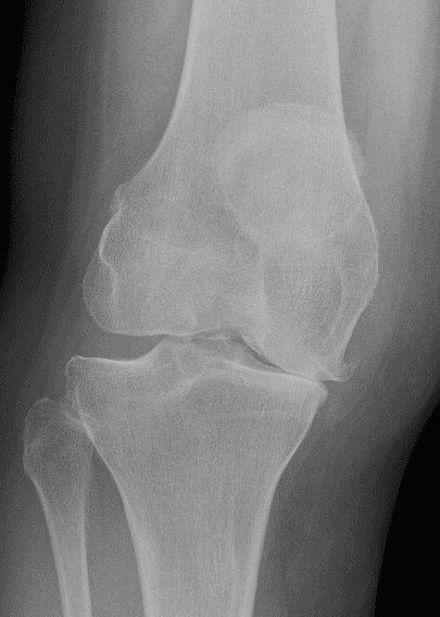
Narrowed joint space due to tissue degradation
Knee arthritis is a common cause of pain, stiffness and reduced mobility. This page explains how it may affect the knee, how it is assessed and the treatment options that may be considered.
Knee arthritis is a clinical condition characterised by the gradual loss or damage of the smooth articular cartilage lining the joint surfaces. As the cartilage thins and wears, the underlying bone surfaces can experience friction, narrowing the joint space. This can lead to joint stiffness, pain, and reduced function. Wording is cautious: symptoms vary greatly between individuals, and X-ray findings do not always correlate with symptom severity.
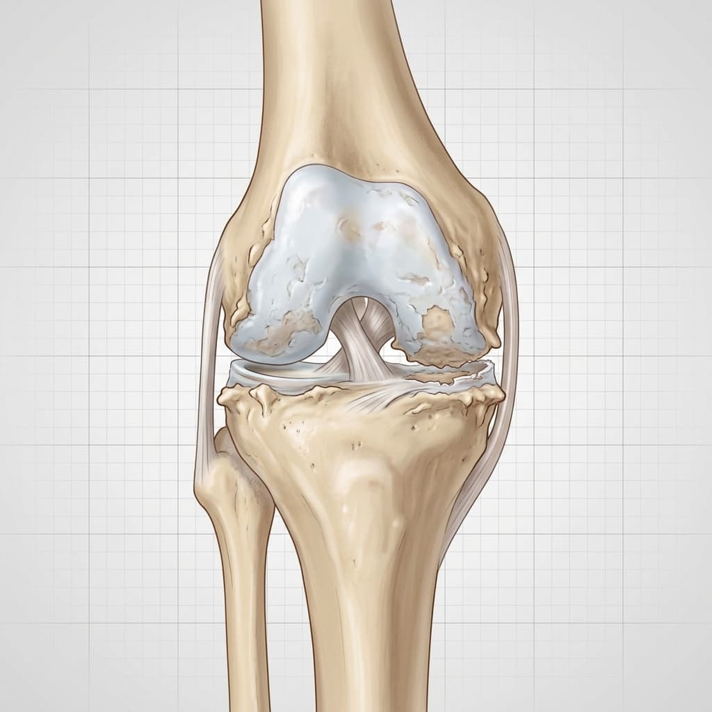
Thinning and erosion of the protective articular cartilage.
Bony projections forming along the joint margins in response to load changes.
Reduced clearance between femur and tibia due to cartilage loss.
Thinning and erosion of the protective articular cartilage.
Bony projections forming along the joint margins in response to load changes.
Reduced clearance between femur and tibia due to cartilage loss.
Understanding structural differences caused by knee arthritis.
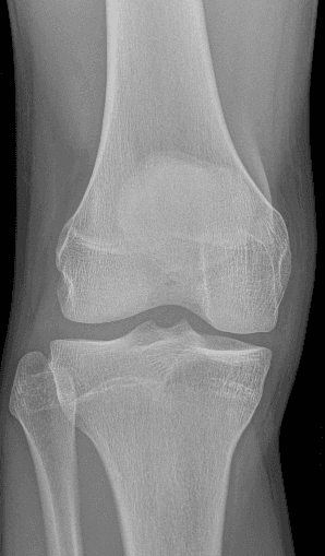 Normal X-rayClinical Reference
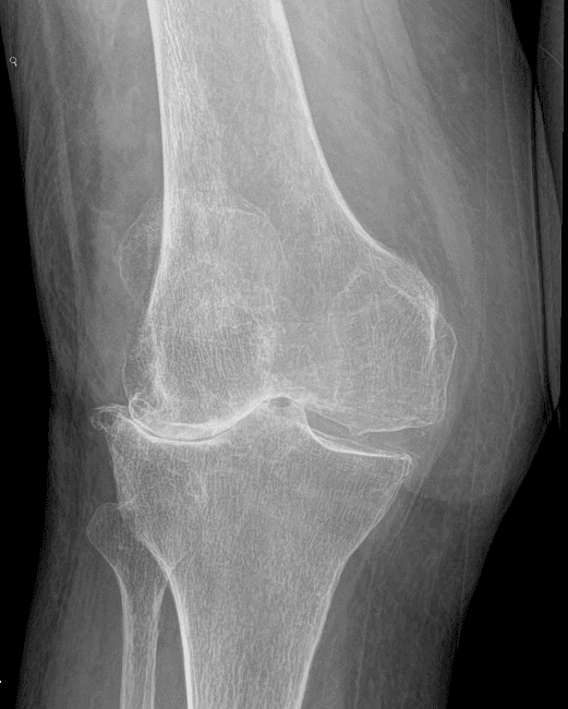 Arthritic X-rayClinical Reference
Normal Anatomy
Wide, even clearance between bone surfaces
Pathological / Affected Anatomy
Narrowed joint space due to tissue degradation
Normal Anatomy
Smooth articular cartilage and strong stabilizing bands
Pathological / Affected Anatomy
Eroded surfaces, osteophytes (bone spurs), or exposed subchondral bone
| Anatomical Feature | Normal Anatomy | Pathological / Affected Anatomy |
|---|---|---|
| Joint Space | Wide, even clearance between bone surfaces | Narrowed joint space due to tissue degradation |
| Structural Integrity | Smooth articular cartilage and strong stabilizing bands | Eroded surfaces, osteophytes (bone spurs), or exposed subchondral bone |
Select any numbered hotspot on the anatomy book illustration below to view details. Use the category filters to simplify the view.
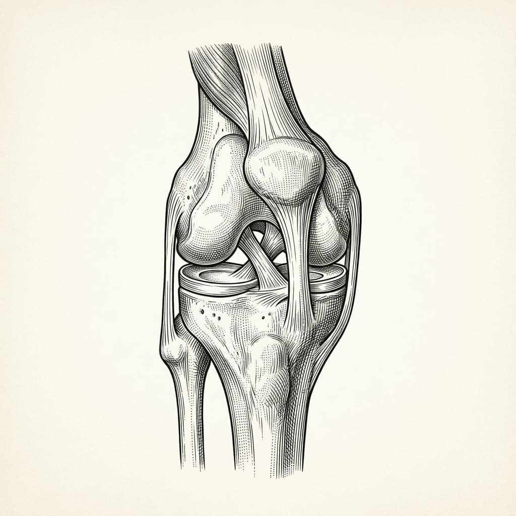
Select a numbered hotspot on the illustration or use the Prev/Next buttons below to browse.
The lower end of the femur forms the upper part of the knee joint. The rounded medial and lateral condyles glide and pivot on the tibial plateau.
A triangular sesamoid bone embedded in the quadriceps and patellar tendon complex. It glides through the trochlear groove, protecting the joint front and multiplying quadriceps leverage.
The main weight-bearing shin bone. The flat tibial plateau forms the lower surface of the knee joint, carrying the menisci.
The smaller outer shin bone. It does not carry joint weight directly but serves as an attachment point for the lateral collateral ligament (LCL) and biceps femoris muscle.
Smooth, wear-resistant hyaline cartilage that caps the ends of the femur and tibia. It provides an extremely low-friction surface, distributing weight and protecting subchondral bone.
A thick, C-shaped fibrocartilage shock absorber on the inner side of the knee joint. It spreads load, improves bone congruency, and aids joint stability.
A circular fibrocartilage shock absorber on the outer side. It is slightly more mobile than the medial meniscus, assisting load transmission across the lateral joint compartment.
A key stabilizing ligament running diagonally deep inside the joint. It prevents the tibia from sliding forward on the femur and controls knee pivoting rotation.
The strongest ligament inside the knee, crossing behind the ACL. It prevents the tibia from sliding backward on the femur.
A strong fibrous band connecting the patella bottom to the tibial tuberosity on the shin bone, transmitting quadriceps extension force.
The thick tendon connecting the quadriceps muscles to the top of the patella. Essential for extending the knee joint.
Symptoms vary, and similar symptoms can occur with other knee conditions. Commonly reported signs include:
The development or onset of this condition can be associated with several mechanical, biological, and lifestyle factors, including:
A comprehensive clinical assessment is performed to understand the root cause of your symptoms and outline appropriate management options.
Imaging and other investigations may be recommended depending on clinical presentation. Common assessments include:
Indicated where clinically appropriate following assessment.
if diagnostic uncertainty or concurrent soft-tissue pathology exists
to rule out inflammatory arthritis in selected cases
Indicates loss of cartilage loading surface on the inner side of the knee.
Increased bone density (whitening) under the worn cartilage zone.
Indicates loss of cartilage loading surface on the inner side of the knee.
Increased bone density (whitening) under the worn cartilage zone.
We offer structured, staged treatment pathways, focusing on conservative and non-surgical approaches. Suitability depends on individual assessment.
Indicated where clinically appropriate.
Explore Treatment >Indicated where clinically appropriate.
Explore Treatment >Indicated where clinically appropriate.
Explore Treatment >Indicated where clinically appropriate.
Explore Treatment >Surgical intervention may be discussed when non-operative treatments do not provide sufficient relief, symptoms significantly restrict daily function or quality of life, and clinical and imaging findings correlate.
unicompartmental) for single-compartment wea
Consultant orthopaedic procedure.
Decisions depend on a detailed diagnosis, symptom severity, patient goals, activity expectations, and general health history. We support shared decision-making, ensuring you fully understand the expected benefits, risks, and alternatives of each pathway.
A consultation can help clarify the cause of your symptoms and discuss appropriate treatment options.
Face-to-face appointments are arranged through the reception team at the chosen clinic. Video consultations use Google Calendar where available.
Treatment pathways range from physiotherapy and bracing to injections or surgical procedures, tailored to your symptoms, diagnosis, and physical goals.
Get notified about new content on Knee Arthritis
We'll only email you about updates on this specific topic — unsubscribe anytime.
This website provides educational and general information about knee symptoms, conditions, and treatments. The content is for informational purposes only and does not constitute medical advice, diagnosis, or a treatment plan. It should not be used as a substitute for a professional consultation, examination, or clinical decision-making by a qualified orthopaedic specialist.
If you are experiencing symptoms or are concerned about a knee problem, please arrange a clinical consultation. If you have urgent symptoms, severe pain, or are unable to put weight on your leg, please read our urgent advice guidance or contact emergency services immediately (dial 999 or NHS 111).